
- Mass Spectrometry for Energy Transformation and Astrochemistry
- Extreme Ultraviolet Electronic Structure Characterization and Lithography
- Electronic Structure Characterization for Operando Micro/Nano Devices
- High-sensitive, Space-Resolved and Time-Resolved Electron Spin Dynamics
- In-situ/Operando Soft X-ray Spectroscopy and Scattering
- Soft X-ray Ptychographic Nanoscopy
- Resonant Coherent Scattering
- High Throughput In-situ/Operando Tender X-ray Spectroscopy
- Tender X-ray Spectromicroscopy and Ptychography
- Test Beamline

Experimental Technique
Ptychographic Coherent Diffraction Imaging (Ptychography)
Scanning transmission x-ray microscopy (STXM)
Low-energy x-ray fluorescence (LE-XRF)
Spectro-microscopy
TEM-compatible imaging
Beamline optics
Overview
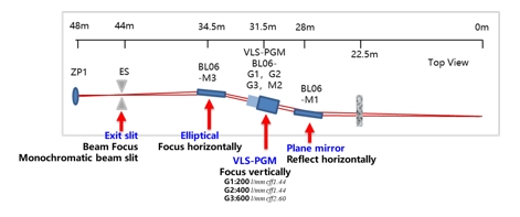
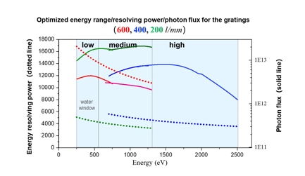
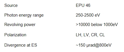
The beamline is sourced by an elliptically polarizing undulator (EPU46). A plane mirror (M1) collects the diverging beam from the undulator and deflects it by 2° to reduce heat load and filter out high energy X-rays. The monochromator is a Plane Grating Monochromator (PGM), consisting a plane mirror (M2) and three different variable-line-spacing (VLS) gratings optimized for varied energy ranges, revolving powers and photon fluxes. The monochromator energy-disperses the X-ray, which is then focused vertically by the VLS gratings. The monochromatized X-ray beam is horizontal focused the ellipsoidal mirror (M3) to the exist slit. At the endstation, the X-ray beam is refocused using a zone plate (ZP) to some specific size (such as 25, 45, 70, 100 nm) for the experiments.
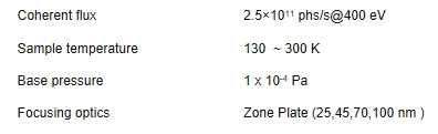
Key Performance
Experimental Endstation
Key Performance
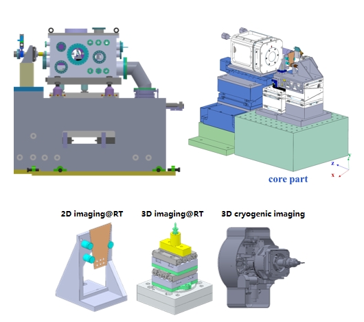
Overview
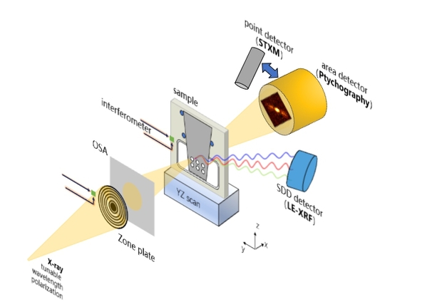
An endstation designed for soft X-ray spectro-nanoscopy, integrates multiple techniques and operational modes, incorporating piezoelectric stages and a laser interferometer.
Techniques: Ptychography, STXM, LE-XRF
Operation modes: 2D/3D imaging; TEM compatible 3D cryogenic imaging
Detectors: sCMOS, PD/APD, 4 channel-SDD, channeltron
Science
To investigate the inhomogeneous distribution of chemical elements with different valence states, and to reveal non-uniform orientations of electron bonds and spins.
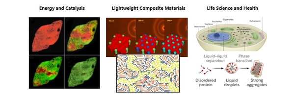
Scope1:
To capture static and dynamic images of ion transfer and valence evolution in energy and catalysis processes, such as the dynamic evolution of the electrode-electrolyte interface layer in all-solid-state batteries during fabrication and operation.
Scope2:
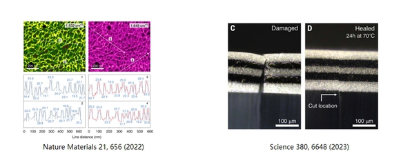
To perform tomography on composite materials composed by light elements using spectral imaging with element-, valence- or polarization-dependent X-ray resonant scattering. Examples include analyzing the morphology of semiconductor hetero-junctions in organic photovoltaic, artificial skin, etc.
Scope3:
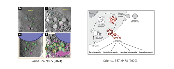
To enable high-contrast and high-resolution imaging of frozen biological cells within the soft X-ray water window. Multimodal imaging combined with SEM, TEM and Super-Resolution Fluorescence Imaging.
People

Useful Link
Related beamlines:
https://sm.lightsource.ca/
https://als.lbl.gov/beamlines/7-0-1-2/
https://www.maxiv.lu.se/beamlines-accelerators/beamlines/softimax/
https://www.helmholtz-berlin.de/pubbin/igama_output?modus=einzel&sprache=en&gid=1884
https://www.elettra.eu/elettra-beamlines/twinmic.html
https://www.diamond.ac.uk/Instruments/Imaging-and-Microscopy/I08.html