
- Mass Spectrometry for Energy Transformation and Astrochemistry
- Extreme Ultraviolet Electronic Structure Characterization and Lithography
- Electronic Structure Characterization for Operando Micro/Nano Devices
- High-sensitive, Space-Resolved and Time-Resolved Electron Spin Dynamics
- In-situ/Operando Soft X-ray Spectroscopy and Scattering
- Soft X-ray Ptychographic Nanoscopy
- Resonant Coherent Scattering
- High Throughput In-situ/Operando Tender X-ray Spectroscopy
- Tender X-ray Spectromicroscopy and Ptychography
- Test Beamline

Experimental Technique
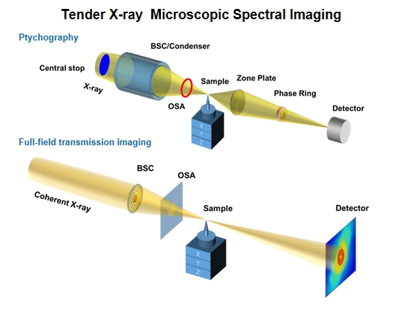
The Tender X-ray Microscopic Spectral Imaging beamline is optimized for the tender X-ray energy range and features both ptychographic and full-field transmission imaging modes. It provides a powerful detection and imaging tool for addressing challenging and critically needed scientific issues in areas such as semiconductor chips, biology, and energy sciences.
Beamline optics
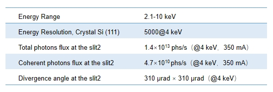
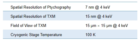
Key Performance
Overview
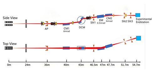
After the beam is exit from the sawtooth wall, a white light slit is set to limit the divergence angle of the beam incident on the monochromator. It is then collimated by a front-end mirror, with a source divergence angle of 30-40 μrad. A liquid-nitrogen-cooled double-crystal monochromator is placed 43 m away from the source point, where the full width at half maximum (FWHM) beam size at the monochromator position is approximately 1.5 mm. The double-crystal monochromator uses Si(111) crystals to meet the requirements for the energy range and energy resolution. To cover the K-absorption edge of phosphorus, the energy range is selected from 2.1 to 10.0 keV. An offset of 20 mm is chosen. Finally, two vertically and horizontally arranged cylindrical mirrors focus the beam at the exit slit2 to generate a secondary source point. The focused beam exhibits nearly equal divergence in the horizontal and vertical directions, forming a defined illumination spot at the entrance of the optical components in the experimental endstation.
Experimental Endstation
Key Performance
Overview
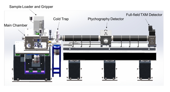
The Tender X-ray Microscopic Spectral Imaging beamline has two imaging systems, ptychography and full-field TXM system, which can carry out three-dimensional nanostructure imaging and spectroscopic imaging.
I. Sample Loader and Gripper
Up to 4 samples can be loaded at a time and maintained in a cryogenic state
II. Ptychography System
Hybrid photon counting X-ray detectors;
High-resolution lensless scanning imaging
III. Full-field Transmission X-ray Microscopy System
Scintillator optically coupled to a dedicated sCMOS;
3D nanostructure imaging and spectroscopic imaging base on X-ray optics such as zone plate
Science
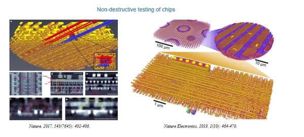
Scientific Scope 1:Ptychography has been applied to the research and detection of chip structures. This technology can reconstruct the internal structure based on the diffracted data of the chip structure without damage, which can be used to find deviations between chip manufacturing and design and discover possible hardware backdoors in the chip.
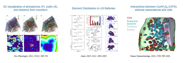
Scientific Scope 2: The energy range of BL09 spans 2.1–10 keV, encompassing the K-absorption edges of elements such as phosphorus, sulfur, chlorine, calcium, titanium, and manganese—elements closely related to environmental pollution, energy catalysis, agricultural ecology, and life sciences. Spectroscopic microimaging techniques can reveal three-dimensional structural changes in samples, along with the spatial distribution and valence state evolution of these elements.
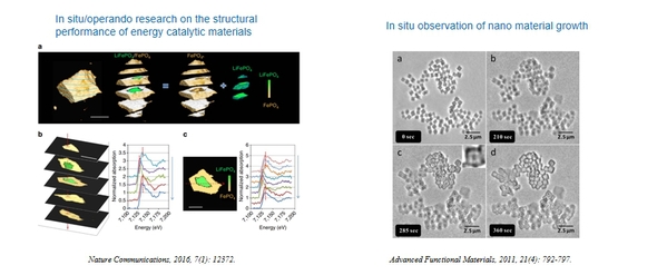
Scientific Scope 3:The high-brightness characteristics of the fourth-generation diffraction-limited ring light source can significantly enhance imaging temporal resolution, enabling in-situ/operando microspectroscopic imaging of batteries and research into nanomaterial growth.
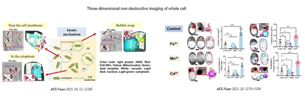
Scientific Scope 4:Cellular fibrosis is a core mechanism of aging and related diseases , involving dysregulation of various fundamental life activities. Therefore, deciphering the mechanisms of cellular fibrosis will contribute to revealing the fundamental principles of life activities.
Imaging technology of BL09 enables cross-scale (10 nm - 10 µm), in situ, and three-dimensional non-destructive cellular imaging. It allows multi-scale (molecular-subcellular structure-cellular) observation, deciphering the intricate interaction networks among biomacromolecules, metabolites, subcellular structures, and other multi-scale, multi-system components.
People
Useful Link
Related beamlines:
Ptychography beamlines:
https://www.psi.ch/en/sls/csaxs
https://www.aps.anl.gov/Microscopy/Beamlines/2-ID-D
https://www.maxiv.lu.se/beamlines-accelerators/beamlines/nanomax/
Full-field transmitted tender X-ray imaging beamlines:
https://www.bnl.gov/nsls2/beamlines/beamline.php?r=18-ID
https://www-ssrl.slac.stanford.edu/content/experimental-stations/bl6-2c
http://e-ssrf.sari.ac.cn/beamlines_2024/sr_72267/beamline_maps/bl18b/xzjs/
https://english.ihep.cas.cn/heps/fa/bl/202112/t20211222_295082.html