

Useful Information
1、Project and Experiment Applications
Before submitting an experimental project, please read the station introduction and research content carefully, or contact the relevant staff of the station to confirm that the project you applied for is suitable for this station. Please register on the Chinese Academy of Sciences Major Scientific and Technological Infrastructure Sharing Service Platform and submit your project application. After the project is approved and the project time is allocated, submit the experiment application on the same platform. The experimental machine time must not exceed the whole project time.
2、Operation Plan and Status
Users can get the operation plan and current status from website, and apply for experimental time reasonably based on their own experimental progress.
3、Preparation Before Experiments
After the user confirms the time of experiments, he can contact the user office staff (yhb@ustc.edu.cn, 0551-63602018) to consult about completing radiation safety training, radiation dose cards, user passes and accommodation.
Users should first carefully read the beamline station introduction or user information page to check the available beamline station equipment or experimental cautions. If there are other matters that need to be understood or consulted, please contact the beamline station staff in time to obtain more relevant information.
Before confirming the experimental arrangements, it is recommended that users communicate with the beamline station researchers to discuss the feasibility of the experiment, preparations, and experimental operation procedures.
For mass spectrometry experiments, users need to confirm the following information with beamline station researchers:
1. For in-situ thermal catalytic reactions, confirm the temperature, pressure, atmosphere and the special gases must be booked in advance to avoid wasting test time;
2. For the in-situ photocatalytic reactions, confirm the requirement of a light source of a specific wavelength, the conversion rate and selectivity of the reaction, the amount of catalyst required, and the feasibility of the experiment;
3. For pyrolysis experiments of solid fuel, confirm the required temperature, gas condition, the ionization energy of target species;
4. For normal test, confirm the suitability of sample phase for the beamline equipment;
5. For mass spectrometry imaging experiments, confirm the sample size, required spatial resolution and characteristics of interested analytes;
6. Other relevant information of experiments.
4、Introduction of Research Content
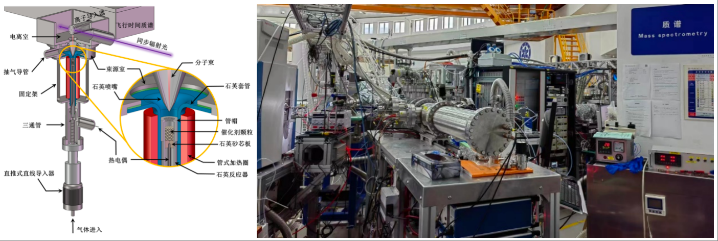
Photoionization mass spectrometry is an experimental technique that accurately characterizes molecules through the mass-to-charge ratio of ions and the photoionization efficiency curve. Synchrotron radiation light sources have the characteristics of high light flux, good stability, and continuously adjustable wavelength, making them ideal light sources for photoionization mass spectrometry. Synchrotron radiation-based photoionization mass spectrometry, combined with molecular beam sampling, is one of the most critical experimental techniques for studying intermediates in gas-solid catalytic reactions, and plays a vital role in the study of energy catalytic reaction mechanisms and catalyst design. In addition, photoionization has the characteristics of strong resistance to matrix effects and no ionization polarity discrimination, and is gradually playing an important role in the simultaneous visualization analysis of polar and non-polar compounds in biological tissues.
Principle of Photoionization Mass Spectrometry
Mass spectrometry (MS) is an important experimental technique for identifying molecular structures. Its basic principle is to ionize the components in the sample in the ion source to generate charged ions with different charge-to-mass ratios to form an ion beam. When a pulse voltage is applied to the acceleration zone plate, the ions are accelerated along the time-of-flight mass spectrometer direction and move toward the detector. When flying through the acceleration zone, the ions gain a certain amount of kinetic energy and enter the free flight zone. Since the ions have the same kinetic energy after acceleration, the ions with smaller mass have a faster speed and arrive at the detector earlier, while the ions with larger mass have a slower speed and arrive at the detector later. Ions with different mass-to-charge ratios are recorded and detected by the detector, and then a mass spectrum is generated. This method can not only provide molecular weight information sensitively and accurately, but also determine the molecular formula by measuring the exact mass formula.
For gases and semi-volatile substances, the traditional electron bombardment ionization is the most widely used source. However, electron bombardment ionization will break down molecule structure. Therefore, for the qualitative and quantitative analysis of complex samples, chromatography separation must be used, which means the impossibility of in situ analysis. Compared with traditional electron bombardment ionization sources, vacuum ultraviolet photoionization can minimize the generation of fragment ions, which is critical for the analysis of multi-component complex systems. According to the experimental requirements, capillary sampling or molecular beam sampling can be selected. The advantage of capillary sampling is that the experimental operation steps are simple and universal, and the reaction process can be monitored online in real time. Molecular beam sampling can cool and preserve active intermediates to detect free radical intermediates in catalytic reactions.
For the non-volatile substances on the solid surface, microdroplets or lasers can be used for desorption. Combined with post-photoionization, the sensitivity of various substances can be increased by 2-3 orders of magnitude, achieving simultaneous analysis of polar and non-polar compounds and improving the coverage of the measured species. Base on a two-dimensional moving stage, the post-photoionization mass spectrometry imaging device can obtain the spatial distribution information of various substances. Mass spectrometry imaging of biological tissues can in situ obtain the spatial distribution information of various metabolites, which is of great significance for studying the biological metabolic mechanism and has broad application prospects in biomedicine, pharmacy, toxicology and other fields.
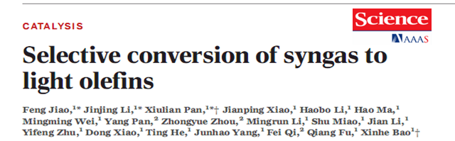
Capture of intermediates during catalytic reactions
About 80% production process of chemical products requires the participation of catalysts. The study of the catalytic reaction mechanism is of great significance for the design of catalysts and the optimization of process conditions. In order to reveal the catalytic reaction process, it is necessary to capture the gas-phase active intermediates of the catalytic reaction in addition to the in-situ characterization of catalysts. Synchrotron radiation photoionization mass spectrometry combined with low pressure catalytic reactor can achieve in-situ characterization of gas-phase intermediates, such as the detection of ketene in syngas to olefins and free radicals in methane activation conversion.
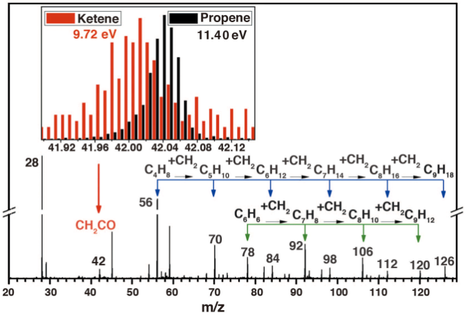
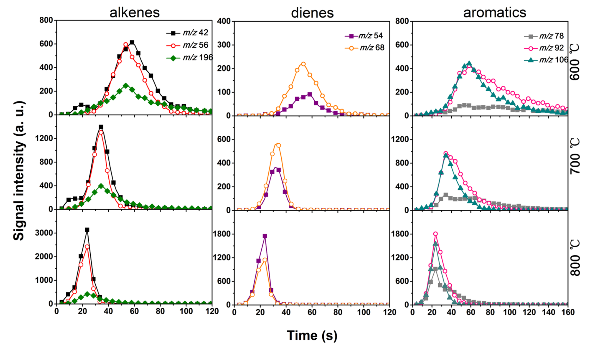
Universal Mass Spectrometry
Capillary sampling based on synchrotron radiation photoionization mass spectrometry is a mass spectrometry analysis method for general samples. It is simple to operate and suitable for sample analysis in various occasions. Two common application scenarios are introduced below: one is the element tracking of trace isotope samples, and the other is the online monitoring of solid pyrolysis reactions. Mass spectrometry is the only analytical technology used for isotope detection and tracking. The amount of products in some photocatalytic reactions is extremely small, and conventional electron bombardment ionization is prone to produce fragments. The high sensitivity and soft ionization characteristics of synchrotron radiation photoionization mass spectrometry can be used to characterize the isotope information in trace gas phase products. Coal and biomass are typical solid raw materials, and their pyrolysis can produce higher-value chemicals. In addition, organic solid waste can also be recycled through pyrolysis. Online characterization of the reaction process using photoionization mass spectrometry is of great significance for revealing its pyrolysis mechanism.
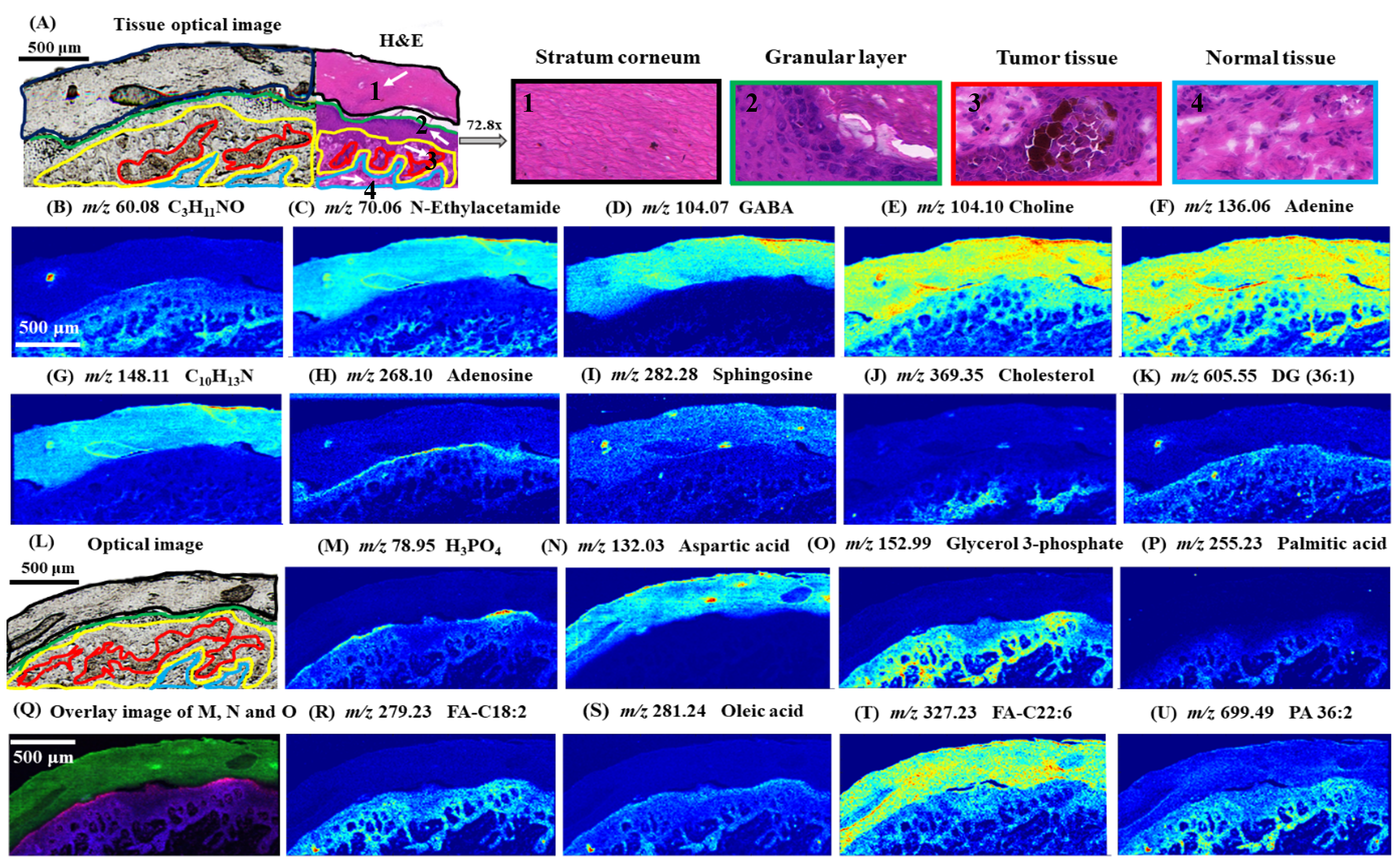
Mass Spectrometry Imaging
Mass spectrometry imaging is a new molecular imaging technology developed based on mass spectrometry. By directly scanning biological samples, the spatial distribution characteristics of multiple molecules can be obtained simultaneously. It has the advantages of no fluorescent labeling and no need for complex sample pretreatment. It has become one of the key technologies in medicine, pharmacy, microbiology and other research fields. At present, the mass spectrometry imaging technology of the mass spectrometry beamline station can analyze samples including animal and plant tissue sections or plant leaves. Both polar and non-polar compounds can be detected, and the spatial resolution can reach up to 4μm. It should be noted that mass spectrometry imaging can not only be used for the visualization of metabolites in biological tissues, but also for research in archaeology, geology and other fields.
Back