

Useful Information
1、Project and Experiment Application
Before submitting an project proposal, please carefully read the beamline introduction and research content, or contact relevant beamline personnel to confirm that the proposed project is suitable for this beamline. Please register on the Chinese Academy of Sciences Major Science and Technology Infrastructure Sharing Service Platform and submit your project application. Once the project is approved and beamtime is allocated, submit your experiment application on the same platform. The experiment duration must not exceed the total allocated beamtime.
2、Beam Supply Schedule and Operational Status
Users can query the beam supply schedule and current operational status to reasonably apply for experimental beamtime based on their experimental progress.
3、Pre-Experiment Preparation
Once the experimental beamtime is confirmed, users can contact the user office staff (yhb@ustc.edu.cn, 0551-63602018) to inquire about completing radiation safety training, obtaining a radiation dose badge, user access passes, and accommodation arrangements.
Users should first carefully read the beamline introduction or user information page to review the available beamline equipment and experimental considerations. If there are any other inquiries or information needed, please promptly contact the beamline personnel for further assistance.
Before confirming the experimental arrangements, it is advisable for users to communicate with beamline researchers to discuss the feasibility of the experiment, preparatory work, and operational procedures.
For experiments at the Infrared spectroscopy and microspectroscopy beamline, users need to confirm the following information with the beamline operations personnel:
1、Confirm the properties (stability, corrosiveness, toxicity, etc.) of the samples being tested, as well as any gases, liquids, and chemical reagents being brought. Gas cylinders must comply with safety regulations, and experiments involving flammable, toxic, or hazardous gases require special communication.
2、For non-in situ tests, confirm the dimensions of the samples, the required spatial resolution for measurement, the energy range of the samples, the measurement modes (transmission, reflection, angle variation, polarization), and the properties of the samples (toxicity, stability when exposed to the atmosphere).
3、For in situ tests, in addition to confirming the relevant information for non-in situ testing, it is necessary to confirm the conditions for testing such as temperature, illumination, atmosphere, and liquids, as well as whether the in situ cell is compatible with the beamline optics.
4、For experiments requiring infrared windows such as ZnSe and CaF2, users should prepare spare windows in advance to prevent disruptions to experiments due to window breakage.
5、Plan the schedule reasonably and prepare spare samples adequately to improve beamtime utilization and avoid wastage.
6、Any other relevant information related to the experiment and the beamline.
7、Users from institutions outside the University of Science and Technology of China must contact beamline staff at least two days prior to the experiment for campus entry reporting.
4、Research Content of the Beamline
The Infrared spectroscopy and microspectroscopy beamline is based on a high-brightness synchrotron radiation source, developing various spectroscopic techniques under conditions such as microspectroscopy imaging, extreme environments (low temperature, high pressure, and strong magnetic fields), and in situ conditions. Research conducted at this beamline encompasses quantum materials, metal-insulator transitions, the optical properties of materials, in situ chemical reactions (photoelectrocatalysis), polymer materials, biological materials, the composition and structure of biological tissues and cells, as well as studies in geoscience, environmental science, paleontology, and archaeology.
Principle of Infrared Spectroscopy
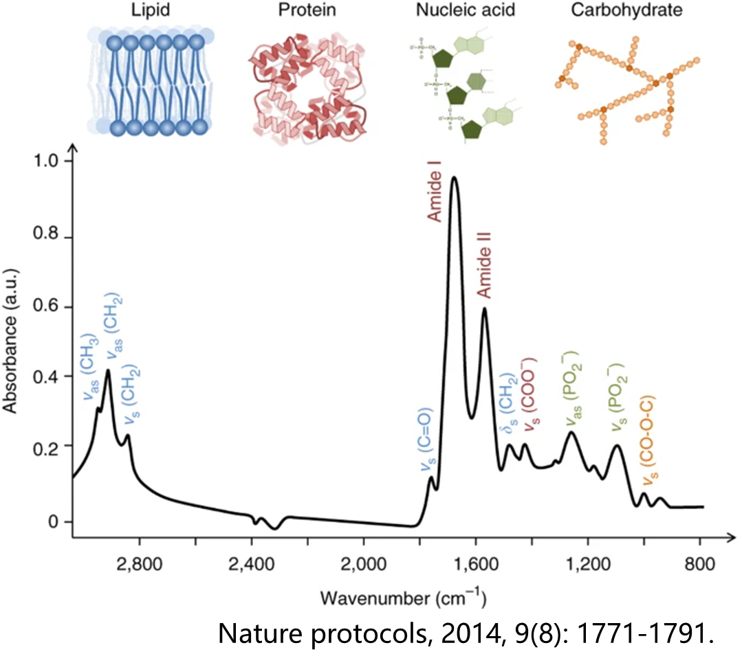
The core principle of infrared spectroscopy is derived from the selective absorption of electromagnetic waves by molecules and the mechanisms of their energy level transitions. According to quantum mechanics, when the energy of an incident photon (E=hν) precisely matches the energy difference between specific vibrational or rotational energy levels within a molecule, the molecule absorbs that photon and undergoes a quantized energy transition. This process must satisfy strict selection rules: only when molecular vibrations or rotations lead to a transient change in the dipole moment does the molecule exhibit infrared activity. For example, the vibrations of polar chemical bonds (such as C=O and O-H) significantly alter the molecular dipole distribution, resulting in strong absorption peaks in the infrared spectrum; conversely, highly symmetrical nonpolar bonds (such as O₂ and N₂) have minimal infrared activity due to their near-zero changes in dipole moment.
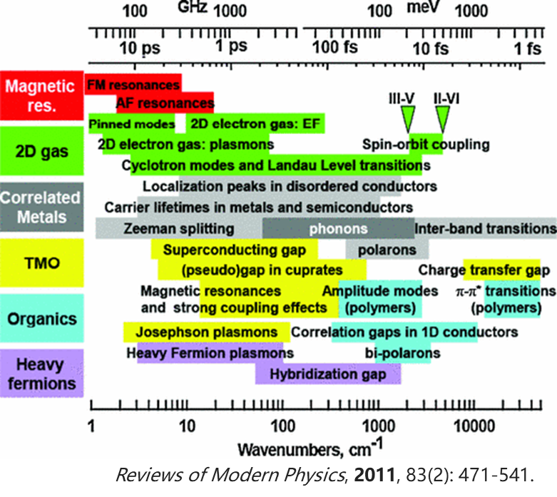
Infrared light typically refers to electromagnetic waves with wavelengths ranging from 0.78 to 1000 μm (corresponding to wavenumbers of approximately 12800 to 10 cm⁻¹), with the mid-infrared region (4000 to 400 cm⁻¹) serving as the core analysis band due to its coverage of the fundamental vibrational energy levels of most chemical bonds. The internal vibrational modes of molecules can be broadly classified into two categories: stretching vibrations (periodic changes in bond length, such as symmetric and asymmetric stretching) and bending vibrations (changes in bond angles, such as in-plane rocking and out-of-plane twisting). The frequencies of these vibrations are determined by the bond strength (force constant k) and the reduced mass (μ) of the bonded atoms, which can be approximately described using the simple harmonic oscillator model (ν=![]() ). For example, the C-H bond, due to the small mass of the hydrogen atom and high bond strength, exhibits stretching vibrations in the high wavenumber range of 2800–3300 cm⁻¹, while the C-Cl bond, with a larger chlorine atom mass, has a significantly reduced vibrational frequency of around 600–800 cm⁻¹.
). For example, the C-H bond, due to the small mass of the hydrogen atom and high bond strength, exhibits stretching vibrations in the high wavenumber range of 2800–3300 cm⁻¹, while the C-Cl bond, with a larger chlorine atom mass, has a significantly reduced vibrational frequency of around 600–800 cm⁻¹.
Through infrared spectroscopy, fingerprint information about molecular structures can be obtained. The positions of characteristic absorption peaks are directly related to functional groups: for instance, O-H stretching vibrations occur at 3200–3600 cm⁻¹, C=O stretching vibrations at 1650–1900 cm⁻¹, and the fingerprint region from 1500 to 400 cm⁻¹ reflects the vibrational coupling of the overall molecular backbone. Additionally, changes in peak shape (such as hydrogen bonding causing broadening of the O-H peak) and shifts in peak position (such as the conjugation effect lowering the C=O vibrational frequency) can reveal information about intermolecular interactions and the influence of the chemical environment. Furthermore, electronic state information such as Drude absorption, superconducting energy gaps, and excitons may also be reflected in the corresponding infrared spectra.
Quantum materials
The energy scales of the rich physical phenomena found in quantum materials are located within the infrared region. The wide-band, variable-temperature infrared spectroscopy (both reflection and transmission) provided by the Infrared Spectroscopy and Microspectroscopy Beamline is essential for investigating crucial information regarding the electronic, phononic, polaritonic, and carrier dynamics, as well as the band structure of quantum materials. This capability enables researchers to gain insights into the fundamental interactions and behaviors within these materials, facilitating advancements in the understanding and application of quantum phenomena.
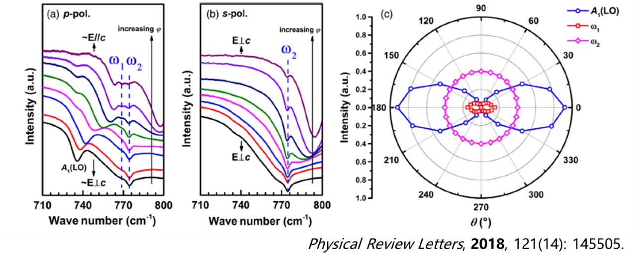
Carbon doping is crucial for the performance of semi-insulating gallium nitride (GaN) in electronic devices. However, the properties of carbon-doped GaN, particularly the lattice site occupancy, have not been thoroughly investigated for a long time. Through polarized Fourier-transform infrared spectroscopy, two localized vibrational modes associated with carbon in semi-insulating GaN were successfully observed. This provided direct evidence of carbon impurities substituting for nitrogen sites in GaN. Additionally, first-principles calculations were employed to validate these findings, effectively resolving a longstanding contentious issue in the field.
In situ infrared spectroscopy captures intermediates
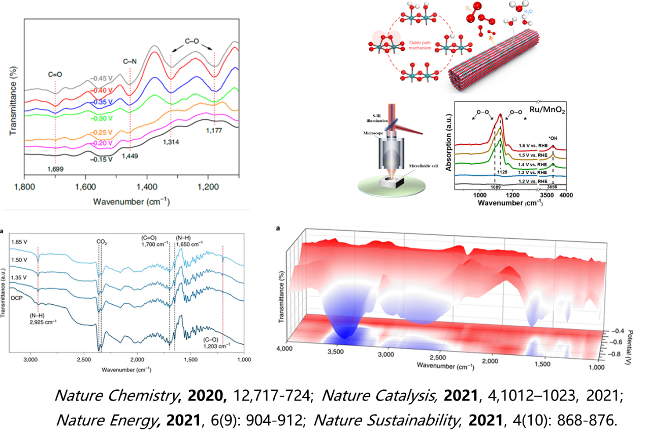
In electrocatalytic reactions, the intermediates formed at the interface between the catalyst surface and the electrolyte (such as adsorbed species like CO, OH, and *OOH) are critical factors that determine the reaction pathway, selectivity, and efficiency. However, these intermediates often exhibit characteristics such as low concentration and spatially localized distribution, making it challenging for traditional non-in situ characterization methods to accurately capture and analyze them under operational conditions (such as applied potential, sustained current, and dynamic mass transfer environments).
Synchrotron infrared microspectroscopy technology offers a groundbreaking solution to this challenge. On one hand, the high brightness and broad spectral coverage of synchrotron radiation sources (ranging from mid-infrared to far-infrared) significantly enhance detection sensitivity, allowing for the detection of surface-adsorbed species at the single-molecule level. On the other hand, microscale focusing techniques enable in situ dynamic monitoring of the working electrode surface, directly capturing the kinetics of the generation and consumption of intermediates during electrochemical polarization processes.
Furthermore, by integrating density functional theory (DFT) calculations to identify the spectral peak assignments, a closed-loop analytical framework can be established that connects structure-activity-mechanism. This multimodal in situ analytical strategy not only deepens the understanding of the dynamic reconstruction processes of electrocatalytic active sites but also provides mechanism-driven theoretical support for the rational design of efficient catalysts.
Infrared spectroscopy under extreme conditions
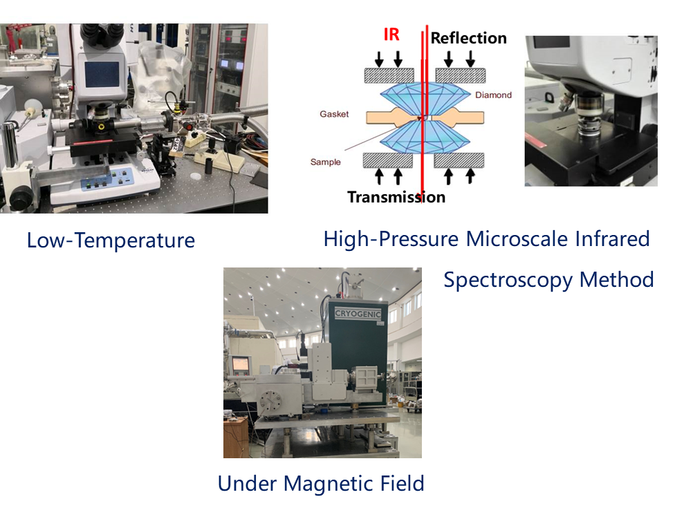
The high brightness advantage of synchrotron radiation is particularly well-suited for conducting spectroscopic measurements in confined spaces, such as under high pressure and strong magnetic fields. The Infrared Spectroscopy and Microspectroscopy Beamline, in conjunction with diamond anvil cells and high-field magnets, has established capabilities for high-pressure infrared spectroscopy and infrared techniques under magnetic fields.
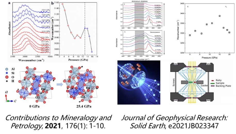 Synchrotron-based high-pressure infrared spectroscopy has opened a new dimension for microscopic dynamic detection in geological research. For instance, in the study of deep Earth materials, this technology can simulate high-pressure environments akin to those found in the mantle (on the order of tens of thousands of atmospheres) and enables the in situ tracking of lattice vibrational modes and the evolution of hydrogen bonding networks in hydrous minerals, such as olivine and wadsleyite, during compression. This provides critical evidence for understanding the storage mechanisms of hydrogen in the forms of hydroxyl groups and molecular water, thereby illuminating the Earth's internal water cycle and the geochemical behavior of hydrogen.
Furthermore, in exploring the nitrogen cycle within the Earth's crust, this technique allows for precise analysis of the diffusion dynamics of ammonium ions (NH₄⁺) within feldspar mineral lattices. By examining the frequency shifts and intensity changes of the NH₄⁺ infrared characteristic peaks under high-pressure conditions, researchers can elucidate the distribution and transport behavior of nitrogen elements in the crust.
Compared to traditional characterization methods, the high brightness and broad spectral coverage of synchrotron radiation, combined with diamond anvil cell (DAC) high-pressure apparatus, enable high spatial resolution detection of material structure and chemical bond evolution under extreme conditions. Such research not only deepens the understanding of the elemental cycling mechanisms between the Earth's spheres but also provides unique technical support for interdisciplinary fields such as planetary science and energy geology.
Materials Science
The Infrared Spectroscopy and Microspectroscopy Beamline integrates wide-band (THz to far-infrared to near-infrared) spectral detection technologies, providing multidimensional research capabilities for condensed matter physics and materials science. Its core advantages lie in the ability to analyze the lattice dynamics of dielectric materials through high-resolution vibrational spectroscopy (such as polariton excitation and phonon mode evolution), establishing quantitative correlations between phonon-electron coupling effects and the frequency response characteristics of dielectric constants. Additionally, by combining the spatial resolution capabilities of microscale imaging (reaching the sub-micrometer level), it is possible to perform in situ characterization of two-dimensional materials (such as graphene and transition metal dichalcogenides), revealing phenomena such as anomalous optical absorption, plasmonic resonance effects, and the electromagnetic wave modulation mechanisms of artificial metamaterials (including metasurfaces and photonic crystals). This powerful experimental support further aids in uncovering quantum effects in low-dimensional systems and in the development of novel optoelectronic devices.
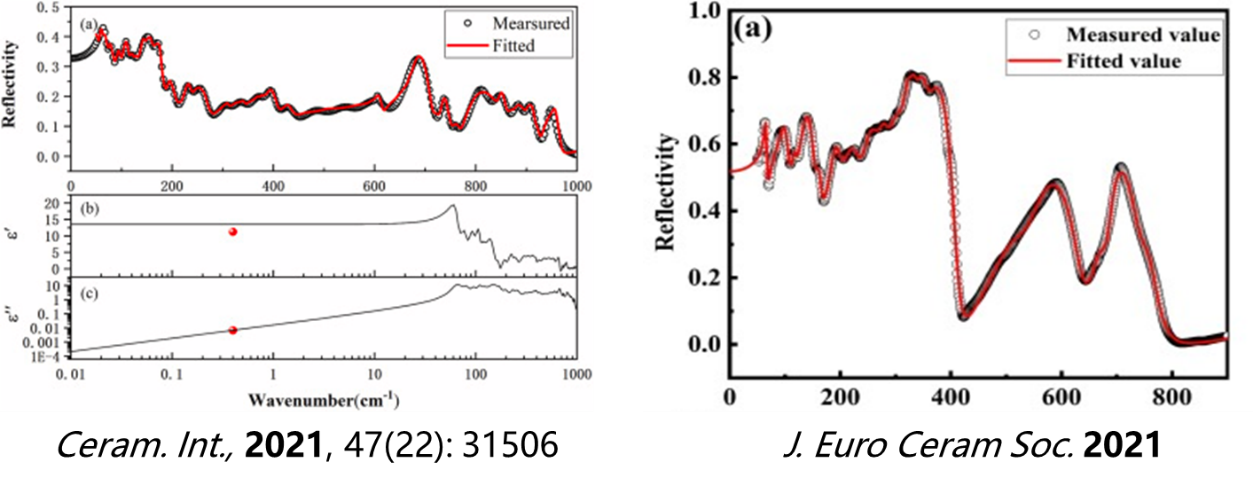
Taking advantage of high-brightness synchrotron wide-band infrared reflection spectroscopy, a systematic study is conducted on the temperature dependence and doping control effects of infrared phonon modes in microwave dielectric materials. This research reveals the microscopic coupling mechanisms between lattice vibrations and dielectric polarization responses, thereby establishing quantitative correlations between key parameters such as dielectric loss and resonance frequency with lattice dynamics. This work provides theoretical guidance for designing low-loss, high-stability 5G devices and promotes the performance optimization of high-frequency communication devices.
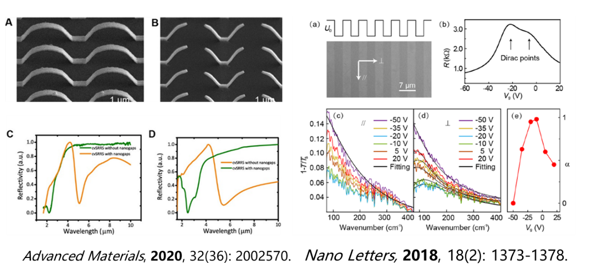
Microscopic infrared spectroscopy, with its diffraction-limited spatial resolution, enables precise analysis of the localized electromagnetic field distribution and optical response characteristics of three-dimensional micro-nano superstructures. This capability provides theoretical support for the design of high-sensitivity infrared detectors and biosensors. Additionally, the combination of polarization-modulated terahertz-far-infrared spectroscopy with in situ electric field manipulation allows for the dynamic tracking of the anisotropic evolution of plasmonic resonance modes in atomically thin graphene ribbons. This research reveals the mechanisms by which carrier concentration and quantum confinement effects modulate terahertz waves, which is of significant importance for the development of plasmonic devices.
Polymer Science
Infrared microspectroscopy imaging has a wide range of applications in polymer science, allowing for the investigation of the local composition, orientation, and conformation of polymer chains, as well as interfacial properties in phase-separated copolymers and composites. The high brightness of synchrotron radiation enables the rapid acquisition of high spatial resolution spectra, as well as time-resolved measurements of dynamic properties under external influences such as mechanical strain, temperature, and chemical conditions.
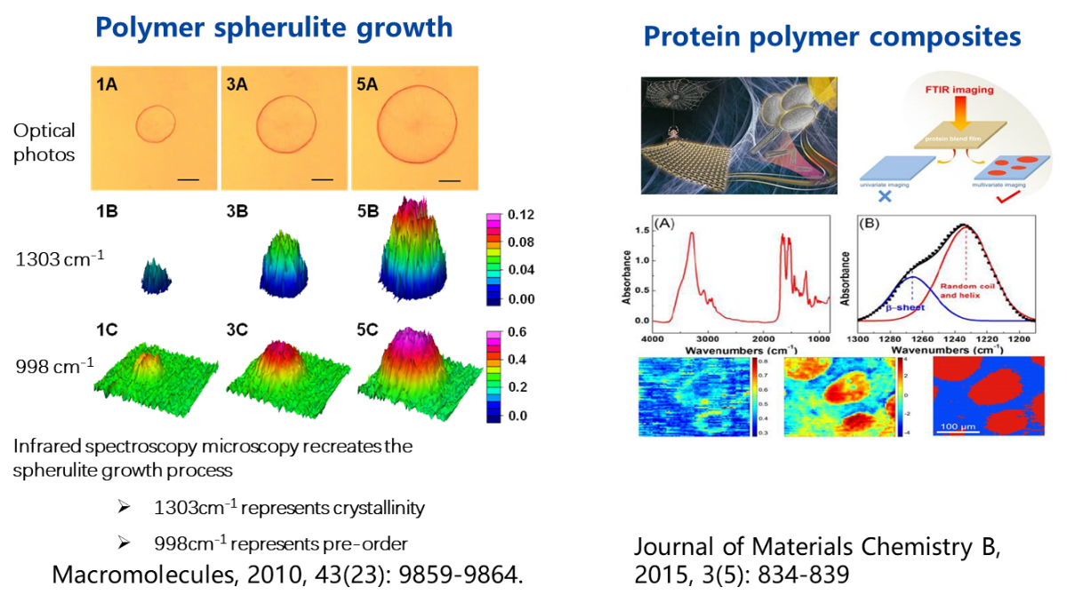
By utilizing synchrotron microspectroscopy imaging technology, in situ dynamic detection of the growth process of polymers can be achieved. For instance, real-time analysis of the infrared spectral responses during the evolution of crystalline regions in polymer spherulite growth can be conducted, combined with external field manipulations to elucidate the crystallization kinetics mechanisms. Furthermore, related techniques can also be employed to investigate the phase behavior of protein-polymer composites in applications such as biomaterials, food industry, and water treatment.
Infrared spectroscopy encompasses a wealth of molecular information related to life. High-resolution infrared microspectroscopy and imaging techniques based on high-brightness synchrotron radiation sources are highly effective methods for studying the molecular composition and structure within biological and生命 systems. These techniques enable high spatial resolution microspectroscopic imaging of single cells and tissues, allowing for the acquisition of molecular composition and structural information at the subcellular level.
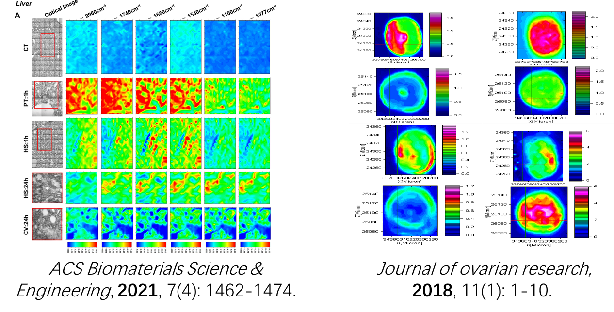
Synchrotron infrared spectroscopic imaging technology, with its diffraction-limited spatial resolution, reveals the dynamic distribution of biomolecules. For instance, it can track the distribution of polyethylene glycol-coated gold nanoparticles in the liver, spleen, and kidneys of mice, quantifying their metabolic pathways through vibrational peaks and elucidating the targeting efficiency of nanomedicine. Additionally, it can analyze the abnormal changes in the secondary structure of keratin in the hair of gastric cancer patients, facilitating the identification of gastric cancer-specific biomarkers and providing new strategies for non-invasive early diagnosis. This technology advances the innovative development of nanomedicine optimization and cancer screening techniques through the precise correlation of molecular fingerprints and spatial distribution.
Back